
FUCCI (Fluorescent Ubiquitination-based Cell Cycle Indicator) is a set of fluorescent probes which enables the visualisation of cell cycle progression in living cells. FUCCI utilises the phase-dependent nature of replication licensing factors Cdt1 and Geminin. A fusion protein of a fragment of Cdt1 (amino acids 30-120) with the fluorescent protein monomeric Kusabira-Orange 2 (mKO2) serves as an indicator of the G1 phase. A fusion protein of a fragment of Geminin (amino acids 1-110 or 1-60) with the fluorescent protein monomeric Azami-Green 1 (mAG1) visualises the S, G2 and M phases. In summary, FUCCI utilises the highly selective, rapid degradation of the replication licensing factors mediated by the ubiquitin proteasome system to give excellent visualisations of the cell cycle.
Cdt1: Cdc10 dependent transcript 1 is a conserved replication factor required for licensing the chromosome for a single course of DNA synthesis. Abundantly expressed throughout the cell cycle, Cdt1 is ubiquitinated by the ubiqutin ligase complex SCFskp2 during S and G2 phases and degraded by proteasome.
Geminin: Geminin inhibits the licensing activity of Cdt1. Geminin interferes with the binding of licensing factors to the replication origin once a chromosome has started to replicate during the S phase. During M and G1 phases, Geminin is ubiquitinated by the E3 ligase complex APCcdh1 and degraded by the proteasome.
What makes FUCCI a powerful research tool?
- Real-time visualisation of cell cycle progression
- Spatio-temporal imaging of cell cycle dynamics
- Utilisation of fluorescent proteins Green mAG1 and Orange mKO2
- Fucci-G1 Orange labels G1 phase nuclei in orange. Fucci-S/G2/M Green labels S/G2/M phases nuclei in green.
By visualising the cell cycle, Fucci is a powerful tool to investigate any process that has to do with cell growth and differentiation, such as the development and regeneration of organs as well as carcinogenesis.
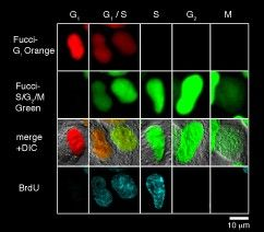
Each cell cycle of the G1, G1/S, S, G2, and M phases can be determined by the combination of Fucci-G1 Orange, Fucci-S/G2/M Green, and an antibody against PCNA (Proliferation Cell Nuclear Antigen). The G1 phase is indicated by orange. Both orange and green were observed in the G1/S phase. Additional immunostaining colour by PCNA was observed at the initiation of the S phase. Cells with pure green fluorescence were either in the S or G2 phase and were distinguished by immunostaining of the S phase. The rest of the cells were classified into the M phase. These results are consistent with the fact that Cdt1 accumulates in G1, while Geminin accumulates in S/G2/M. (Provided by Dr. Sakaue-Sawano at RIKEN. Cell (2008) 132:487-98).
Applications
- High Content Imaging – FUCCI can be used for the visualisation and analysis of single-cell metrics which can provide rich data for analysis.
- Cancer Inhibitor Screening – FUCCI was used to test the effect of selinexor on cancer cell growth. After imaging the cell cycle effects of selinexor using FUCCI, it was determined that G-1 or early S-phase treated cells show the strongest response and most often die or arrest.
Originally posted on: https://resources.mblintl.com/learning-center/research-area/fluorescent-proteins/cell-cycle-indicator/?hsCtaTracking=2b726f5e-2439-4d0c-9ac3-6c5761d3a54a%7Cdb77aea6-4980-4393-8e5e-164964f382a2
Caltag Medsystems is the distributor of MBL products in the UK and Ireland. If you have any questions about these products, please contact us.