
Background
KACTUS has developed two transmembrane protein display platforms: virus-like particles (VLPs) and nanodiscs. These platforms are designed for functional display of multipass transmembrane proteins in their native conformation. KACTUS produce these full-length membrane proteins using detergent-free extraction methods, which preserve the structural integrity and biological activity of the proteins. Their products are suitable for a range of applications including immunisation, ELISA, yeast display, flow cytometry, and SPR. For these applications, choose from unlabelled, biotinylated, or fluorescent proteins. Additionally, their transmembrane proteins undergo bioactivity and purity testing using SPR, ELISA, flow cytometry, and HPLC. This ensures functional performance and batch consistency for reproducible results. Researchers can apply these products to antibody discovery, antibody screening, analytical method development, and immunisation.
Membrane Protein Technology Platforms
Virus-Like Particle (VLP)
Copolymer Nanodisc
Mammalian-expressed transmembrane proteins displayed on VLPs for native conformation and boosted immunogenicity
Transmembrane proteins expressed on membrane-like structures composed of phospholipids and copolymers
Virus-like Particle (VLP) Proteins
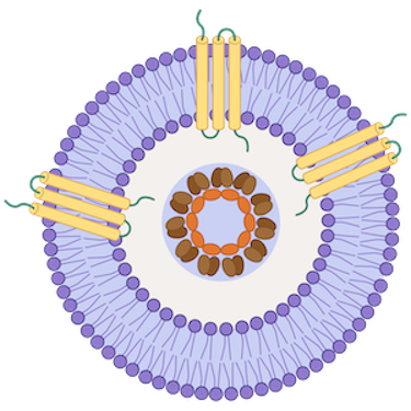
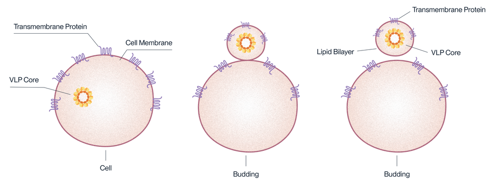
Virus-like particles (VLPs) are small nanoparticles composed of virus shell or capsid proteins. These proteins self-assemble into virus-like structures, mimicking the organisation and morphology of the actual virus. However, VLPs are absent of viral infectious genomes, making them non-infectious and safe for use in various applications, including antibody immunisation, screening, vaccine development and gene therapy.
Advantages of VLP Proteins
VLPs display the target protein in a dense manner on their surface. This boosts the immune response to the target protein, making VLPs useful for immunisation, especially with antigens that are usually present in low amounts or don’t trigger a strong immune reaction by themselves. VLPs also allow for the soluble expression of multipass transmembrane proteins without the use of detergents.
Applications
- Blood sample determination: ELISA
- In vivo pharmacokinetic analysis
- Antibody immunisation, screening, and functional characterisation
- CMC method development
- Affinity determination via ELISA, SPR
Features
- Site-Specific Biotinylation
- VLP capsid proteins optimised for each protein
- Proven biological activity via ELISA and SPR
- Mammalian cell expression system for natural folding and glycosylation
- Boosted immunogenicity for antibody drug discovery
Product Validation Data
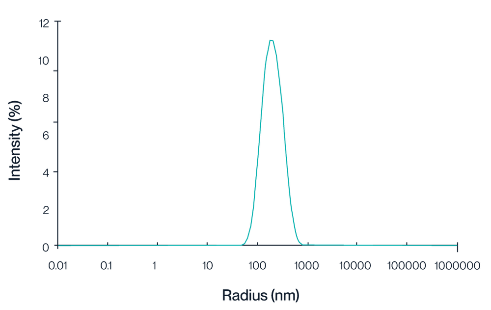
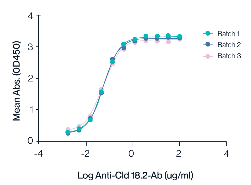
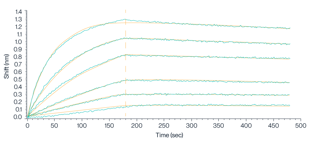
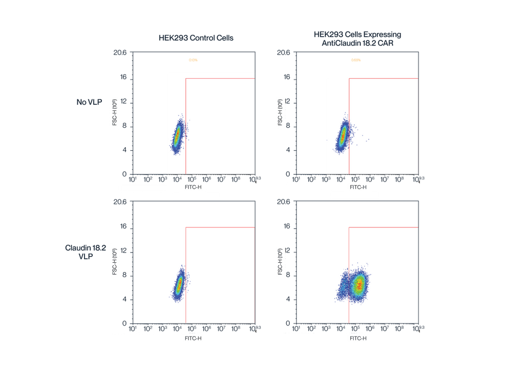
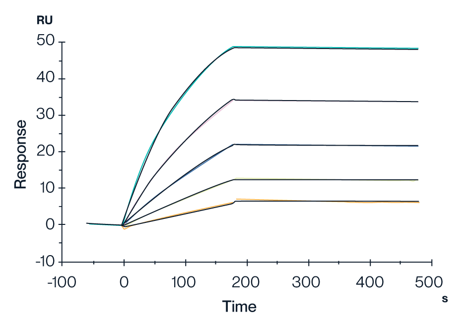
Claudin 18.2 VLP
KACTUS was the first company to express full-length, wild-type Claudin 18.2. This complex 4-transmembrane domain protein has therapeutic potential for gastric and esophageal adenocarcinomas. Despite interest in developing drugs aimed at Claudin 18.2, technical challenges in expressing high-quality Claudin 18.2 protein had limited the potential for antibody drugs around this target. Quality recombinant target proteins are necessary for immunisation, screening, and analytical method development. However, by leveraging virus-like particles, KACTUS successfully produced full-length Claudin 18.2 protein in mammalian cells. Their consistent activity and purity testing results demonstrate the functional integrity and bioactivity of their VLP-displayed protein platform.

Nanodisc Proteins
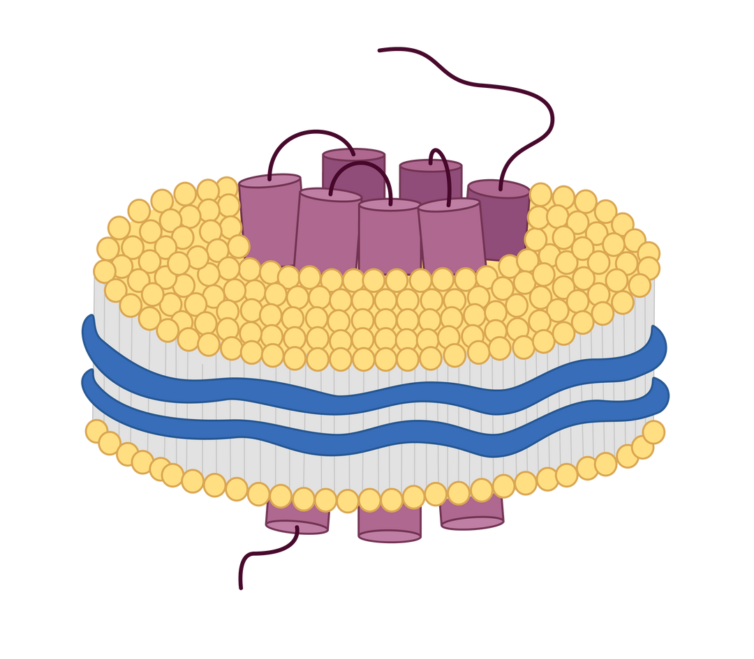
Nanodiscs are small disc-shaped structures that mimic a cell membrane. A target protein is present in a lipid bilayer, which is surrounded by membrane scaffold proteins or copolymers. This discoidal structure enables solubilisation without the use of detergents. At KACTUS, they use a proprietary amphipathic copolymer to ensure performance and reliability. Their nanodiscs can be applied in applications such as ELISA, SPR, BLI, and yeast display.

Advantages of Nanodisc Proteins
Using nanodisc-displayed membrane proteins has several benefits. It maintains the natural conformation of the protein, provides water-soluble proteins, and displays the protein in a native-like environment. Moreover, because nanodiscs can be extracted without detergents, they are superior for membrane proteins that are sensitive to maintaining activity with detergents.
Applications
- Yeast display screening
- Functional in vitro assays
- CAR expression testing
- PK/PD Studies
- Analytical Tests, ELISA, SPR, BLI
Features
- Native protein structure and conformation
- Detergent-free extraction
- Water-soluble
- Proven biological activity via ELISA and SPR
- Mammalian cell expression system for natural folding and glycosylation
Product Validation Data
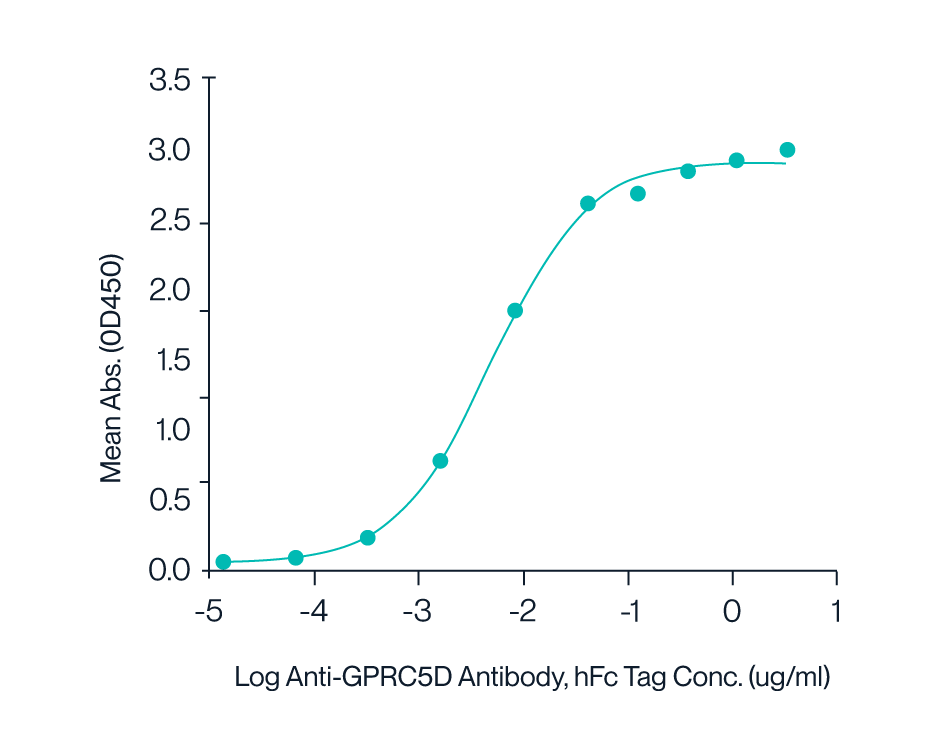
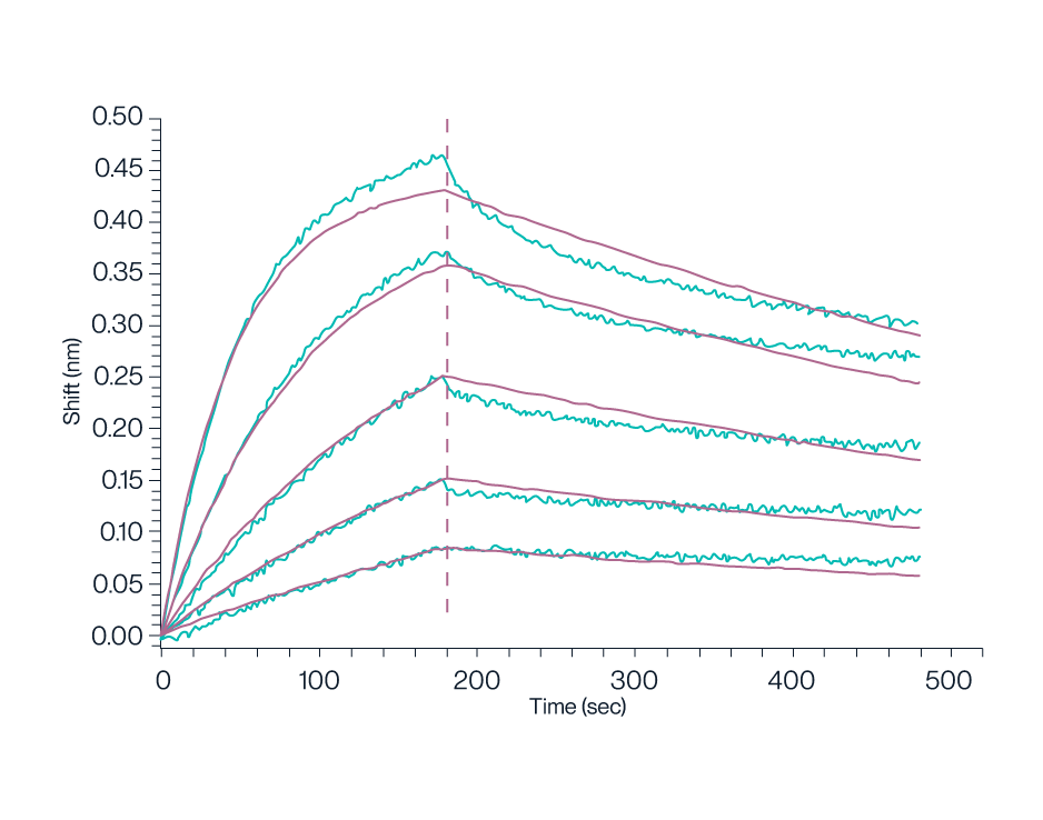
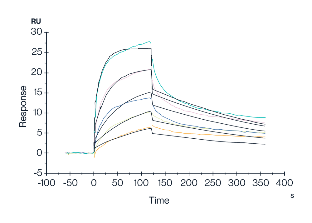
GPRC5D Nanodisc
GPRC5D is a complex, 7-transmembrane domain protein and a popular target for multiple myeloma treatment. This protein is being targeted in CAR-T cell therapy, antibody-drug conjugates, and bispecific antibody drugs (with CD3 protein) for myeloma. To facilitate GPRC5D drug development, KACTUS has developed a full-length GPRC5D nanodisc with verified bioactivity via ELISA and SPR. Along these lines, they have developed biotinylated nanodisc proteins to broaden use cases for this membrane protein. Their biotinylated GPRC5D nanodisc has high binding activity and can be used in analytical method development and pharmacokinetic analysis.

Caltag Medsystems is the distributor of KACTUS products in the UK and Ireland. If you have any questions about these products, please contact us.