MANF Antibody
Product Code:
PSI-4349
PSI-4349
Host Type:
Rabbit
Rabbit
Antibody Isotype:
IgG
IgG
Antibody Clonality:
Polyclonal
Polyclonal
Regulatory Status:
RUO
RUO
Applications:
- Enzyme-Linked Immunosorbent Assay (ELISA)
- Immunofluorescence (IF)
- Immunohistochemistry (IHC)
- Western Blot (WB)
No additional charges, what you see is what you pay! *
| Code | Size | Price |
|---|
| PSI-4349-0.02mg | 0.02mg | £150.00 |
Quantity:
| PSI-4349-0.1mg | 0.1mg | £449.00 |
Quantity:
Prices exclude any Taxes / VAT
Stay in control of your spending. These prices have no additional charges, not even shipping!
* Rare exceptions are clearly labelled (only 0.14% of items!).
* Rare exceptions are clearly labelled (only 0.14% of items!).
Multibuy discounts available! Contact us to find what you can save.
This product comes from: United States.
Typical lead time: 14-21 working days.
Contact us for more accurate information.
Typical lead time: 14-21 working days.
Contact us for more accurate information.
- Further Information
- Documents
- References
- Related Products
- Show All
Further Information
Additional Names:
MANF Antibody: ARP, ARMET, ARP, Mesencephalic astrocyte-derived neurotrophic factor, Arginine-rich protein
Application Note:
WB: 0.125-2 μg/mL; IHC: 2.5 μg/mL; IF: 20 μg/mL.
Antibody validated: Western Blot in human and rat samples; Immunohistochemistry in human samples; Immunofluorescence in human samples. All other applications and species not yet tested.
Antibody validated: Western Blot in human and rat samples; Immunohistochemistry in human samples; Immunofluorescence in human samples. All other applications and species not yet tested.
Background:
MANF Antibody: MANF, also known as ARMET, was initially identified as a protein containing an arginine-rich region that was highly mutated in a variety of tumors. More recently it was identified as a mesencephalic astrocyte-derived neurotrophic factor with selectivity for dopaminergic neurons, similar to glial cell line-derived neurotrophic factor (GDNF) and CDNF. In rat brain slices, MANF enhanced nigral gamma-aminobutyric acid release. Like GDNF and CDNF, MANF has selective neuroprotective activity for dopaminergic neurons suggesting that it may be indicated for the treatment of Parkinson's disease. Expression of MANF has also been shown to be induced during ER stress, suggesting that it may play a role in protein quality control during ER stress.
Background References:
- Shridhar et al. Oncogene 1996; 12:1931-9.
- Shridhar et al. Cancer Res. 1996; 56:5576-8.
- Petrova et al. J. Mol. Neurosci. 2003; 20:173-88.
- Lindholm et al. Nature2007; 448:73-7.
Buffer:
MANF Antibody is supplied in PBS containing 0.02% sodium azide.
Concentration:
1 mg/mL
Conjugate:
Unconjugated
DISCLAIMER:
Optimal dilutions/concentrations should be determined by the end user. The information provided is a guideline for product use. This product is for research use only.
Homology:
Predicted species reactivity based on immunogen sequence: Bovine: (92%)
Immunogen:
Anti-MANF antibody (4349) was raised against a peptide corresponding to 12 amino acids near the amino terminus of human MANF.
The immunogen is located within the first 50 amino acids of MANF.
The immunogen is located within the first 50 amino acids of MANF.
ISOFORMS:
Human MANF has one isoform (182aa, 21kD). Mouse MANF has one isoform (179aa, 20kD) and Rat MANF also has one isoform (179aa, 20kD). 4349 can detect human, mouse and rat.
NCBI Gene ID #:
7873
NCBI Official Name:
mesencephalic astrocyte derived neurotrophic factor
NCBI Official Symbol:
MANF
NCBI Organism:
Homo sapiens
Physical State:
Liquid
PREDICTED MOLECULAR WEIGHT:
Predicted: 20kD
Observed: 26 kD
Observed: 26 kD
Protein Accession #:
P55145
Protein GI Number:
23503040
Purification:
MANF Antibody is affinity chromatography purified via peptide column.
Research Area:
Neuroscience
SPECIFICITY:
This antibody does not cross-react with CDNF.
Swissprot #:
P55145
User NOte:
Optimal dilutions for each application to be determined by the researcher.
VALIDATION:
Recombinant Protein Test (Figure 2): Anti-MANF antibodies (4349) detected human MANF recombinant protein at different concentrations.
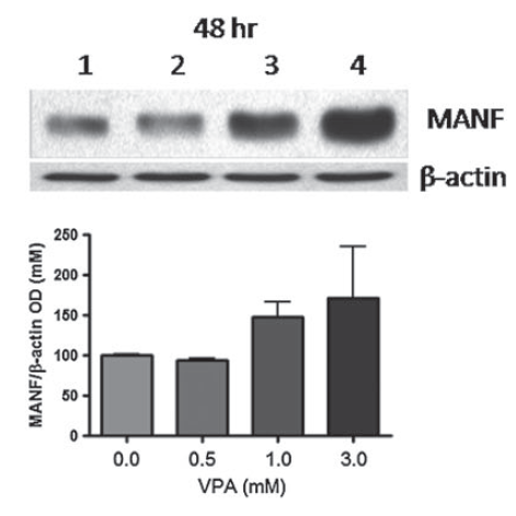
Induced Expression Validation (Figure 6): MANF expression detected by anit-MANF antibodies was up-regulated by VPA treatment.
Documents
References
- Almutawaa et al. Induction of neurotrophic and differentiation factors in neural stem cells by valproic acid. Basic Clin Pharmacol Toxicol. 2014;115(2):216-21. PMID: 24460582
- Niles et al. Valproic acid up-regulates melatonin MT1 and MT2 receptors and neurotrophic factors CDNF and MANF in the rat brain. Int J Neuropsychopharmacol. 2012;15(9):1343-50. PMID: 22243807
- Walaa S. Almutawaa. Induction Of Neurotrophic and Differentiation Genes in Neural Stem Cells by Valproic Acid. McMaster University, M.Sc. Thesis. 2012PMID:
- Merja H. Voutilainen. CDNF and MANF in an experimental model of Parkinson's disease in rats. University of Helsinki, Ph.D. Thesis. 2010PMID:
Related Products
| Product Name | Product Code | Supplier | MANF Peptide | PSI-4349P | ProSci | Summary Details | |||||||||||||||||||||||||||||||||||||||||||||||||||||||||||||||||||||||||||||||||||||||||||||
|---|---|---|---|---|---|---|---|---|---|---|---|---|---|---|---|---|---|---|---|---|---|---|---|---|---|---|---|---|---|---|---|---|---|---|---|---|---|---|---|---|---|---|---|---|---|---|---|---|---|---|---|---|---|---|---|---|---|---|---|---|---|---|---|---|---|---|---|---|---|---|---|---|---|---|---|---|---|---|---|---|---|---|---|---|---|---|---|---|---|---|---|---|---|---|---|---|---|---|---|


